Image source: Rensselaer Polytechnic Institute
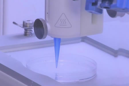
A team of scientist at the Rensselaer Polytechnic Institute have developed a new 3D printing method that uses novel bioinks to create living skin complete with blood vessels that connect to the host system when transplanted.
These artificially created skin grafts represent a significant step forward in 3D bioprinting. While living skin tissue has been 3D printed before that accelerates wound healing, the printed tissue does not have a fully functioning vascular system and eventually dies.
“Right now, whatever is available as a clinical product is more like a fancy Band-Aid. It provides some accelerated wound healing, but eventually it just falls off; it never really integrates with the host cells,” said, Pankaj Karande, PhD, associate professor of chemical and biological engineering, member of the Center for Biotechnology and Interdisciplinary Studies (CBIS), and lead researcher for the study.
Karande and his team have been attempting to 3D print fully functional living skin for several years. They have previously used two types of human cells to create a bioink that can be printed into a skin like structure, but the lack of vasculature has proven to be a problem.
To solve this problem, the researchers created new bioinks that include more cell types and found the 3D printed skin formed a biologically relevant vascular structure within a few weeks. The key was to use a combination of animal collagen and structural cells typically found in skin grafts, along with endothelial cells and human pericyte cells. Endothelial cells form the inside layer of cells in blood vessels and pericyte cells wrap around the endothelial cells.
Two bioinks were developed, the first contained human foreskin dermal fibroblasts, human endothelial cells, and human placental pericytes in rat tail type 1 collagen. This bioink was used to print the dermis. A second bioink was used to print the epidermis layer, which consisted of human foreskin keratinocytes (KCs).
The endothelial cells and pericyte cells assembled to form interconnected microvascular networks and the pericytes appeared to improve maturation of the keratinocytes, which formed an epidermis-like barrier.
The 3D skin they created was grafted onto a mouse and functioned like living skin. The blood vessels in the graft appeared to connect with the blood vessels in the wound bed of the mouse and were perfused within 4 weeks of the graft being transplanted. Since blood was flowing through the skin graft and nutrients were being delivered, the skin graft survived.
Before this method could be used on humans, the researchers would need to use gene editing technology to prevent the skin from being rejected, and they are not yet at that stage. One that issue has been solved, the researchers believe their new skin could be suitable for use on diabetic patients to accelerate the healing of ulcers. Healing is slow in diabetic patients, so small skin grafts could greatly accelerate that process.
The research is detailed in the paper – 3D bioprinting of a vascularized and perfusable skin graft using human keratinocytes, (…) – which was recently published in Tissue Engineering Part A. DOI: 0.1089/ten.TEA.2019.0201