A new imaging technique has been developed by researchers at the University of Toronto that can be used to identify cholesterol in arterial plaque, which could lead to faster diagnosis and treatment of atherosclerosis
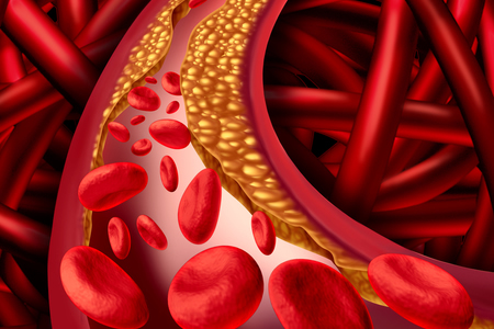
Atherosclerosis is a disease caused when plaque accumulates in the arteries. Plaque is comprised of calcium, fat, cholesterol and other substances. It builds up over time and hardens causing narrowing of the arteries. There are no noticeable symptoms at first, but as the disease progresses it can lead to heart attacks and strokes.
Treatment generally involves lifestyle changes and various medications to reduce blood pressure, cholesterol, and prevent the formation of blood clots. Early intervention is key; however, the disease is largely symptomless in the early stages and half of all men with severe atherosclerosis have no symptoms, so the disease is often diagnosed once it has progressed significantly.
Angiography is the main method used to check for narrowing of the arteries – A form of x-ray using a contrasting agent to show the arteries. While this test can show plaque, it provides no morphological or compositional information and cannot differentiate between different forms of soft plaque.
The researchers use an imaging approach known as photoacoustic radar. It is a combination of hybrid optical-acoustic imaging technology, low-power continuous wave lasers, and frequency-domain signal processing.
The new imaging technique is only sensitive to atherosclerotic plaque lipids. It is not affected by other light absorbers in the artery walls or instrumentation-generated noise signals.
Andreas Mandelis, professor of mechanical and industrial engineering and electrical and computer engineering at the University of Toronto and co-author of the paper said, “When co-registered with intravascular ultrasound (IVUS), [the imaging system] could reveal a complete cross sectional image of lipids in animal coronary arteries and human aorta that delivers accurate location and quantitative radial depth-profiles of atherosclerotic disease.”
There is only one hurdle left to overcome before the imaging system is ready for clinical use. The researchers must develop an ultrasonic transducer that is small enough to fit inside a human coronary artery. The researchers believe that last hurdle can be overcome quickly and the system will be ready for clinical use in about a year.
Further information can be found in the paper – Frequency-domain differential photoacoustic radar: theory and validation for ultrasensitive atherosclerotic plaque imaging – which was recently published in SPIE Journal of Biomedical Optics. DOI: 10.1117/1.JBO.24.6.066003