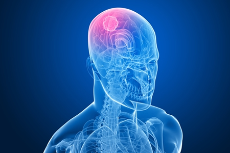
The Mayo Clinic is pioneering the use of new imaging scans that reveal the firmness of brain tumors and the degree to which they are attached to surrounding brain tissue prior to surgery, giving surgeons an accurate picture of the level of difficulty of the surgery and the probability of success.
The Mayo Clinic is the first and only medical center to offer brain magnetic resonance elastography (MRE) imaging and slip interface imaging to patients with brain tumors.
While brain tumors can be identified using other imaging techniques such as MRI scans, neurosurgeons often discover during surgery that tumors are stiffer than expected, making removal more of a challenge. By accurately determining the degree of firmness of the tumor prior to surgery, neurosurgeons can determine the least invasive and most effective method for removing the tumor, for example, sucking the tumor out through the patients nose rather than undergoing a craniotomy, which carries far greater risks.
Slip interface imaging provides neurosurgeons with vital information about the interface between the tumor and the surrounding brain tissue. If tumors are discovered to be more adherent to the surrounding brain tissue during surgery it greatly increases the risk of complications.
In some cases, the tumor may have a capsule-like interface, which makes removal far easier. In other cases, the tumor may be fixed to the surrounding tissue and have a rigid connection. By determining the nature of the interface, neurosurgeons can plan their surgery accordingly, allocating the proper amount of time for the surgery and also determining the safest method of extraction. All of the necessary information can be gathered prior to opening up the patient’s head, which has a major impact on patient safety.
Mayo Clinic neurosurgeon Jamie Van Gompel, M.D., explained the importance of the two imaging techniques saying “Brain magnetic resonance elastography and slip interface imaging help avoid surprises in surgery that could lead to complications for the patient.”
The Mayo Clinic has recently published the results of a slip interface imaging study on 25 patients revealing the level of tumor adhesion. Out of the 25 patients that were involved in the study, the Mayo Clinic was able to determine that 16 individuals had brain tumors that could easily be removed, with nine of the patients found to have tumors with adhesion to the brain. The remaining patients has some brain adhesion. The accuracy of the slip interface imaging scans were confirmed during surgery.
The Mayo Clinic Study – Slip interface imaging based on MR-elastography preoperatively predicts meningioma–brain adhesion – was published in the Journal of Magnetic Resonance Imaging in February, 2017.