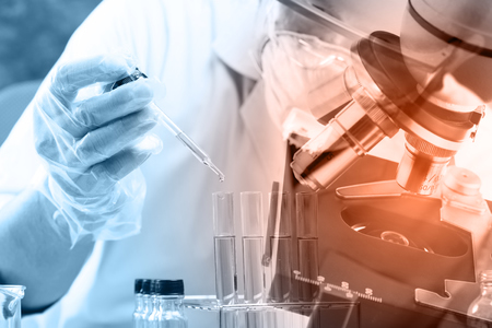
University of Illinois Chicago researchers have developed a new approach for controlling the deposition of hydrogel to direct single cell function. Hydrogels are water-soluble polymers that are used to mimic the extracellular matrix and support cells. Hydrogels are used in research, but also have therapeutic uses, but the current techniques used for depositing the hydrogels lack control.
Most experiments mix cells with large quantities of hydrogel, but this approach may not ideally reflect conditions in the body. This approach also lacks control and makes it hard to study interactions between the cells and their surroundings.
The new method can be used to more accurately replicate conditions in the body, bringing the ratio of hydrogel to cells in line. The researchers found that when cells are suspended in thin hydrogel droplets they expand more rapidly and experience more tension than when they are suspended in bulk gels consisting of the same material.
Using their new approach, the researchers were able to encapsulate a single cell in a tiny droplet of hydrogel, which they showed helped them encourage bone marrow stem cells into specialized cells. The researchers believe the increase in tension encourages stem cells to more readily become bone cells than when bulk gels are used.
“We show that varied gel deposition alone has a profound impact on the rate of isotropic cell volume expansion in the presence of an adhesion ligand, which subsequently regulates membrane tension and stem cell differentiation in a predictable manner,” explained the researchers in their paper. “Results from this study will help facilitate precision engineering of cell–material interactions for fundamental biological science and translational therapeutic applications.”
The researchers explained that their method can be adapted for use with single cell sequencing technologies and can help to understand single cell heterogeneity in biophysical cell–matrix interactions.
Their approach could also be used to augment the osteogenic potential of donor MSCs. The use of a minimal amount of materials could help to reduce the risk of foreign body reaction, as well as decrease the cost of materials.
You can read more about the new technique in the paper – Controlled Deposition of 3D Matrices to Direct Single Cell Functions – which was recently published in the Advanced Science. DOI: 10.1002/advs.202001066