Human mesenchymal stem cells (hMSCs) are present in bone marrow and play an important role in the repair and regeneration of organ damage. These cells travel from the bone marrow and differentiate into the cells needed to repair tissue damage. They also migrate to tumors and interact with cancer cells, but little is known about the effect hMSCs have on cancer cells. There is no consensus of opinion on the role hMSCs play in cancer metastases, with some research indicating hMSCs promote metastasis with other studies suggesting hMSCs reduce metastasis.
Research conducted by Biochemist Mj Brown, a Ph.D student at the School of Sport, Exercise and Health Sciences at Loughborough University, has shed light on interactions between hMSCs and breast cancer cells.
When breast cancer metastasizes and tumors are formed in other parts of the body, termed secondary breast cancer, the outlook for patients is poor. While treatments can be provided to control the disease, there is no cure for secondary breast cancer.
Brown was studying the effect of hMSCs on the breast cancer cell line, MDA-MB-231, which is highly aggressive. She found that hMSCs had a major effect and substantially reduced the invasiveness of the cancer cells.
The research was conducted using a 3-D model of the cancer cells. It is far more common for 2-D models of cancer cells to be used in research. While 2D models are useful, they have severe limitations. Cancer cells in the body are naturally in 3-D, and the cancer cells interact with all cell surfaces that surround them. 3D models allow cancer to be modeled more accurately.
Brown’s supervisor for her PhD and co-author of the study, Dr. Mhairi Morris, is an expert in 3-D cancer modelling. Together they developed a 3-D model of MDA-MB-231 cancer cells and, assisted by Loughborough hMSC expert, Dr. Liz Akam, explored the effect of hMSCs on breast cancer cells.
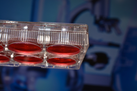
3-D spherical tumors were created by co-culturing them with hMSCs and growing the cell cultures suspended from the lid of a tissue culture dish. After the spheroids had been grown, they were then embedded in a matrix that mimics conditions inside the body. After five days, the invasiveness of the breast cancer cells was measured using the length of the projections formed by the cancer cells in the matrix. Brown found that the hMSCs significantly reduced the length of the projections, thus helping to reduce the invasiveness of the cancer cells.
While establishing the ideal cell culture medium to grow the 3-D spherical tumors, Brown found the source of the serum had a significant effect on growth of the cells. That finding could help other researchers develop other 3-D models of tumors for use in research.
You can read more about the research in the paper – Determining Conditions for Successful Culture of Multi-Cellular 3-D Tumour Spheroids to Investigate the Effect of Mesenchymal Stem Cells on Breast Cancer Cell Invasiveness – which was recently published in the journal Bioengineering. DOI: 10.3390/bioengineering6040101
