Approximately 1 in 60 children in the United States have been diagnosed with autism spectrum disorder (ASD). Obtaining that diagnosis requires a one on one consultation with a qualified clinician and takes between 2 and 4 hours. Even then, the assessments are subjective and are based on the level of experience of the clinician.
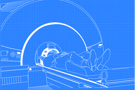
Functional magnetic resonance imaging (fMRI) could serve as a more objective method of diagnosing ASD and could not only improve the accuracy of diagnosing the developmental disorder, but also the speed of getting that diagnosis.
A recent fMRI study on children showed the fMRI method to be both quick and accurate and could lead to the development of the first objective brain-based method of diagnosing ASD.
The study was conducted by researchers at Wake Forest School of Medicine who used fMRI scans to measure the responses of a part of the brain involved in assigning value to social interactions – the ventral medial prefrontal cortex (vmPFC). The children were presented with various environmental cues and the scans showed the vmPFC response.
The study was conducted on 40 children between 6 and 18 years of age, 12 of whom had been diagnosed with ASD and 28 were normally developing children.
While undergoing an fMRI scan, the study participants were shown eight images of objects and people multiple times. Out of the 8 images, two were of a favorite object or person of the child undergoing the test, three were of other objects and three were of other people.
The images were selected from a database widely used in psychological experiments and were either pleasant, neutral, or unpleasant. The fMRI test took between 12 and 15 minutes, after which the children were presented with the same images on a computer screen and were asked to rank them in order, from pleasant to unpleasant. The children were also shown pairs of images and had to say which of the images they preferred.
The researchers found that the responses in the vmPFC were different in each of the two groups of children. It required 30 seconds of fMRI data to be able to differentiate between the two groups, and therefore determine which children had ASD.
“How the brain responded to these pictures is consistent with our hypothesis that the brains of children with autism do not encode the value of social exchange in the same way as typically developing children,” said Kenneth Kishida, Ph.D., assistant professor of physiology and pharmacology at Wake Forest School of Medicine and lead researcher for the study.
Based on this study, Kishida believes that a 30-second fMRI scan would be all that is required to obtain an effective measurement to determine whether the brain responds normally to social and non-social stimuli. The researchers are now studying parts of the brain involved in different aspects of ASD and hope that they will be able to gain an understanding of each child’s position on the spectrum to help develop personalized treatments.
The findings of the study are detailed in the paper – Diminished single-stimulus response in vmPFC to favorite people in children diagnosed with Autism Spectrum Disorder – which was recently published in the journal Biological Psychlogy. DOI: 10.1016/j.biopsycho.2019.04.009