Biomedical researchers in China have constructed artificial ears from human cartilage cells that have been used to correct ear deformities in five children with the congenital ear deformity microtia.
Children born with microtia have an underdeveloped pinna. In severe cases, there is a total absence of the outer ear. The condition often results in impaired hearing due to inefficient conduction of sound to the inner ear. The deformity can result in self-esteem problems, and also makes wearing glasses difficult.
Many individuals with the condition do not have an associated abnormality of the inner ear, so surgical reconstruction of the outer ear can improve hearing. While artificial ears can be constructed and surgically attached, the ultimate goal is to grow ears in the lab for transplantation.
The team of Chinese researchers have done just that. They grew artificial ears from seed cartilage cells harvested from the patients which were grown on a biodegradable scaffold fixed to a 3D printed model of the patient’s ear.
To create the model the researchers took a scan of the patient’s healthy ear and created a digital image using 3DPRO software. The image was then mirrored and the digital image was used as a guide to 3D print a mold on a Z Corp Spectrum 510 3D printer.
The mold was used to cast a model ear out of clay and silicone on which an artificial ear could be grown. The researchers covered the model with a biodegradable mesh scaffold. Microtia Chondrocyte (MCs) cartilage cells were harvested from the patients and were evenly distributed on the scaffold, and the artificial ear was placed in a cell culture solution. After growing the cells for 12 weeks, the model was removed and the artificial ear was surgically attached to the patient with the patients skin draped over the cartilage. Vacuum drainage was used to help fit the skin to the shape of the artificially grown ear.
The treatment was administered to five children aged between 6 and 10. The first patient was assessed over a period of two and a half years and the researcher report there was a “satisfactory aesthetical outcome with mature cartilage formation.” The process was subsequently repeated on a further four patients.

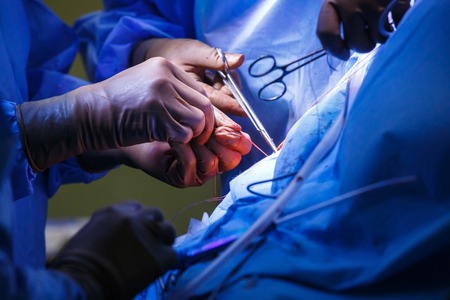
Image Source: G. Shou et al., EBIOMEDICINE
The researchers detail their technique in their paper – In Vitro Regeneration of Patient-specific Ear-shaped Cartilage and Its First Clinical Application for Auricular Reconstruction – Available online through eBiomedicine.